How is VUR diagnosed and graded?

Urinary reflux diagnosis and grading scale

A VUR diagnosis usually comes after a febrile UTI is confirmed

When a child presents with a febrile urinary tract infection (fUTI), it could indicate vesicoureteral reflux (VUR), commonly referred to as urinary reflux.

VUR is most often detected as a result of diagnostic investigations of fUTI or of hydronephrotic (distended) kidney. It may also be suspected in children presenting with high blood pressure or kidney insufficiency.

Diagnosing VUR

Accurate diagnosis and grading of VUR can help with the implementation of individualized treatment plans. Diagnosis requires a micturating cystourethrogram (MCUG), typically performed by a radiologist equipped for the fluoroscopy and catheterization of children.

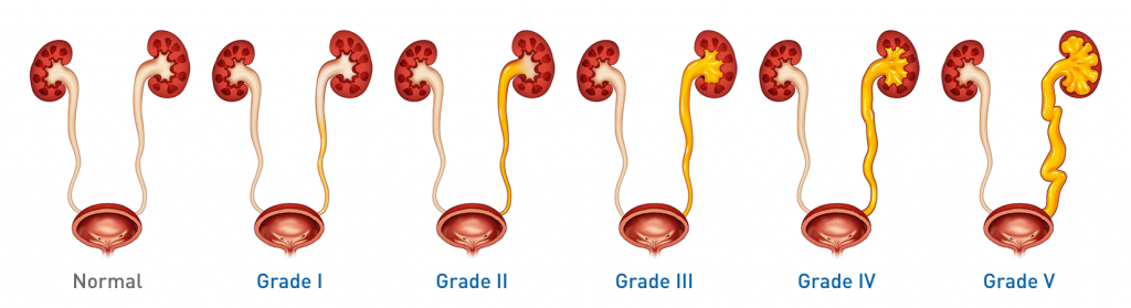
As a result of MCUG assessment, any VUR will be graded from I to V based on the dilation of the ureter and presence and degree of distortion of renal structures.

Imaging Procedures after first UTI per the 2010 American Urologic Association (AUA) guidelines for the management and screening of primary vesicoureteral reflux in children:3

  • Recommendation: Perform a renal and bladder ultrasound (RBUS) to assess the upper urinary tract
  • Option: DMSA (technetium-99m-labeled dimercaptosuccinic acid) renal imaging can be obtained to assess the status of kidneys for scarring and function

If the RBUS is normal, the AUA does not recommend proceeding with a MCUG.

In 2011, the American Academy of Pediatricians presented an update to the clinical practice guidelines for the diagnosis and management of the initial UTI in febrile infants and children 2-24 months. In the updated guidelines, MCUG is only indicated if renal and bladder ultrasonography (RBUS) reveals hydronephrosis, scarring or other findings suggesting either high-grade VUR or obstructive uropathy.4

However, a 2013 study of children with their first UTI and normal RBUS shows that in 24% of patients, VUR would not have been detected.5 The same study shows that 15% of these children had recurrent pyelonephritis and 7% went on to surgical intervention.5

RBUS may not detect VUR after a first UTI5

In another study reviewing patient records from 2002-2004 under the 2011 guidance, it was found that the majority of patients would have gone undiagnosed under the updated AAP guidance:6

  • 17.2% of patients with normal RBUS had renal injury identified on DMSA renal scan
  • 62.1% of patients with normal RBUS had grade III or higher VUR
Grades of VUR

More severe VUR is associated with a lower chance of spontaneous resolution and more severe renal scarring and increased complications7,8

The International Classification System for VUR:9

  • Grade I: Reflux into nondilated ureter
  • Grade II: Reflux into renal pelvis and calyces without dilation
  • Grade III: Reflux with mild to moderate dilation and minimal blunting of fornices
  • Grade IV: Reflux with moderate ureteral tortuosity and dilation of pelvis and calyces
  • Grade V: Reflux with gross dilation of ureter, pelvis, and calyces, loss of papillary impressions, and ureteral tortuosity

Consider referring a patient to a paediatric urologist for the following10:

  • Suspected or confirmed febrile urinary tract infections (fUTIs)
  • Individualized management of VUR
  • Assessment and treatment of related or underlying conditions such as bladder or bowel dysfunction

References:

  1. Elder JS. Vesicoureteral reflux. In: Kliegman R, Nelson WE, eds. Nelson Textbook of Pediatrics. 19th ed. Philadelphia, PA: Elsevier/Saunders; 2011:1834-1838.
  2. Fisher DJ, Steele RW. Pediatric urinary tract infection. Medscape.
  3. American Urological Association. Management and screening of primary vesicoureteral reflux in children: AUA guideline. 2010.
  4. American Academy of Pediatricians. Urinary tract infection: clinical practice guideline for the diagnosis and management of the initial UTI in febrile infants and children 2 to 24 months. Pediatrics. 2011;128(3):595-610. DOI: https://DOI.org/10.1542/peds.2011-1330
  5. Juliano TM, Stephany HA, Clayton DB, et al. Incidence of abnormal imaging and recurrent pyelonephritis after first febrile urinary tract infection in children 2-24 months. J Urol. 2013;190:1505-1510. DOI: 10.1016/j.juro.2013.01.049.
  6. Suson KD, Mathews R. Evaluation of children with urinary tract infection- Impact of the 2011 AAP guidelines on the diagnosis of vesicoureteral reflux using a historical series. J Pediatr Urol. 2014;10:182-185. DOI: https://DOI.org/10.1016/j.jpurol.2013.07.025
  7. González E, Papazyan JP, Girardin E. Impact of vesicoureteral reflux on the size of renal lesions after an episode of acute pyelonephritis. J Urol. 2005;173:571-575. DOI: DOI: 10.1097/01.ju.0000151263.36909.91
  8. Shaikh N, Ewing AL, Bhatnagar S, Hoberman A. Pediatrics. 2010;126:1084-1091. DOI: 10.1542/peds.2010-0685
  9. Lebowitz RL, Olbing H, Parkkulainen KV, Smellie JM, Tamminen-Mobius. International system of radiographic grading of vesicoureteral reflux. Pediatr Radiol. 1985;15:105-109. DOI: 10.1007/BF02388714
  10. Peters CA, Skoog SJ, Arant BS Jr, et al. Summary of the AUA guideline on management of primary vesicoureteral reflux in children. J Urol. 2010;184(3):1134-1144. DOI: 10.1016/j.juro.2010.05.065
X